An introduction to electron micrography, including process and use, and the growing creative applications for this technology.
Electron Micrograph Images
Artistic, creative images of electron micrographs. Images used solely for educational purposes and belong to their respective owners. See "Sources" for information.
Information
Online and text resources for information about electron micrography, both as a science and as an emerging form of art.
Sources
A list of websites from which I gained photographs and information. Bibliography of the available text literature on the subject.
I have a number of different sites. If this is not what you were looking for, try these links:
.the procrastination project.
.completely random.
When people think of electron micrography, the image they usually get in their minds is this:

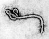
The image on the left is Ebola virus in liver cells by Dr. Tom Geisbert of the Centers for Disease Control. The one on the right is the Ebola Zaire virus, also from the Center for Disease Control (both images are in the public domain).
Compare the right-hand picture of Ebola with the picture featured in the header. The version of Ebola in the header is from an art installation series entitled, "The Art of Death," by Hunter O'Reilly. (Other images from this art installation are used for the other headers on this site). These pieces of art are created by molding plexiglass into the image of famous viruses and then illuminating them brightly with neon, using their electron micrographs as a reference point. Although these images are not directly related to my topic, they are representative of my point: electron micrography is moving out of the domain of the scientific and taking a place in legitimate artistic expression.
Process
Electron micrographs are created with the use of an electron microscope. The development process is similar to traditional photography. But one of the electron microscope's limitations is that even though it can see things as small as a single virus particle, even map it in three dimensions, the optics are such that all the images are in black and white and with a flattened depth of field.
Manipulation as a Route to Art
This is where digitization and digital color come in. I see these manipulated images as being separate from regular scientific photography. Their intent is not to document (since the colors, at times, remove information from the image), but to dazzle.